Location: Groupe de Physique des Matériaux (GPM), Saint Etienne du Rouvray (France) http://gpm.univ-rouen.fr/fr
Duration : 36 months, starting autumn 2026 (ANR funded project DynaMETSAT (ANR-25-CE42-2131-01)
Contact: Williams Lefebvre (williams.lefebvre@univ-rouen.fr)
No microscopy technique today can provide a holistic view of a material. Most recent transmission electron microscopes (TEMs) and scanning transmission electron microscopes (STEMs) easily achieve sub-Angström spatial resolution, while allowing elemental mapping at the same scale. Meanwhile, electron tomography has unambiguously demonstrated the possibility to image atomic positions and defects. In these instruments, some physical properties are now accessible, again with increased resolution. However, as far as an ultimate imaging would allow correlating physical properties with a “perfect” determination of atomic species and atomic positions in 3D, one must recognize that such a tool is not yet available. Aside from electron microscopes, Atom Probe Tomography (APT), is intrinsically a 3D technique which allows reconstructing materials with a sub-nm resolution and a high chemical sensitivity1,2.
APT relies on intense electrostatic fields to remove the atoms from the surface of a needle-shaped specimen (20-50 nm radius at their apex), kept at cryogenic temperature, in the form of ions projected onto a position-sensitive detector. The ions’ time-of-flight is used to retrieve their respective mass-to- charge-state ratio, allowing elemental, and even isotopic identification. The impact position and elemental nature are used, in combination with a projection model, to build a 3D point cloud in which the atoms from the material are repositioned. Ion trajectories follow the electrostatic field lines which are governed by the immediate geometry of the specimen surface at each step of the field evaporation sequence. APT reconstructions hence require prior knowledge or hypotheses about the specimen geometry, since the dynamic evolution of APT specimen surface during field evaporation is not known. Moreover, in heterostructured materials (e.g. quantum wells, transistors, ceramic matrix- composites, batteries…), complex variations of specimen surface due to varying evaporation field condition between phases strongly deteriorate spatial resolution of APT.
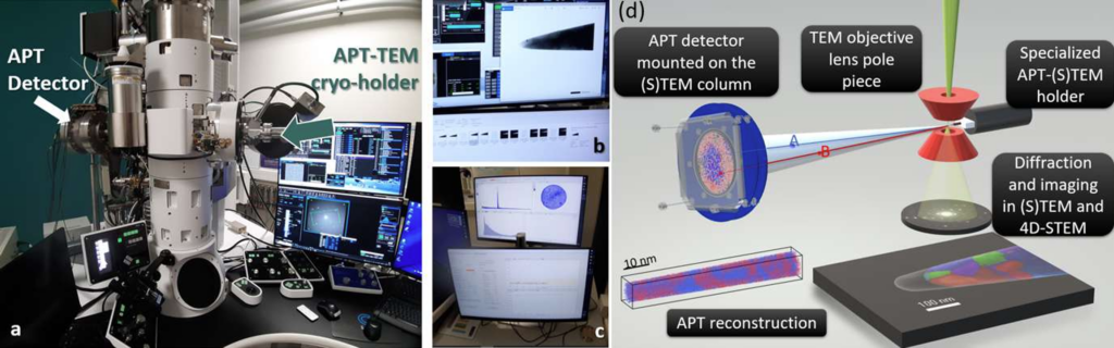
Setup in operation at GPM and designated as SATMET3. (a) SATMET, consisting an a JEOL F200 combined with a customAPT. (b) APT specimen imaged in BF STEM. (c) Acquisition sequence in APT. (d) Sketch illustrating the possibilities offered by SATMET.
In this project (ANR funded DynaMETSAT project), we will reveal the time-resolved geometric evolution of APT specimens and the distribution of electrostatic field at the specimens’ apex with the use of a unique instrument combining an Atom Probe with a (S)TEM3. With this instrument which was designed and implemented by the instrumentation TEM at GPM, we will be able to make the link between the dynamic evolution of specimen surface with the dynamic variations of detection events on the APT detector. In addition, electric field distribution maps at the tip apex will be acquired and compared to simulated distributions calculated for the given specimen geometries determined in TEM. This approach will initiate a paradigm shift in the way APT reconstructions are performed and make a major step toward the high-fidelity 3D microscopy of complex materials of technological interest (e.g. batteries, catalysts), for which the lack of precision of APT reconstructions is until now a bottleneck.
Organization of the work of the PhD candidate: The PhD candidate will be trained to (S)TEM and will follow the internal TEM school at GPM and additional formations. The student will also the Atom probe Tomography school organized every year in November at GPM. The student will become an expert in the use of (S)TEM using various modes (diffraction, TEM, STEM and tomography). The student will conduct the extraction of data obtained by dynamic TEM and APT acquisitions, will use these data to implement modified field evaporation simulations based on those designed by François Vurpillot. Ultimate goal of this PhD work will be to achieve TEM informed APT reconstructions based on dynamic imaging of APT acquisitions in TEM.
Profile: Candidates must hold a MSc in Physics, Materials Science or other related areas. Previous experience in TEM and a background in characterization techniques will be considered, as well a good knowledge in materials-beam interaction. A prior knowledge of APT will be an advantage. Strong organizational skills and ability to work as part of a team shall be attested.
Application: The PhD student will be co-supervised by Prof. Williams Lefebvre and Prof. François Vurpillot. Interested candidates should send a CV, a letter of motivation and the names of 2-3 references to williams.lefebvre@univ-rouen.fr
1. Gault, B., et al. Atom Probe Microscopy. vol. 160 (2012).
2. Williams Lefebvre Ulrikson, François Vurpillot, Xavier Sauvage. Atom Probe Tomography Put Theory Into Practice. (2016).
3. Da Costa, G. et al., Bringing atom probe tomography to transmission electron microscopes. Nat Commun 15, 9870 (2024).